The decision to surgically remove an eye is difficult for both surgeon and patient. This is because of the profound psychological effect of loss of an eye on the patient. This procedure is therefore a last resort.
Removal of the eye may be necessary after severe ocular trauma, to control pain in a blind eye, to treat some intraocular malignancies, in severe eye infections that are unresponsive to medical therapy, and for cosmetic improvement of a disfigured eye1. The choice of procedure to accomplish this is best made by an informed patient.
The methods of surgically removing the eye are evisceration, enucleation and exenteration. Evisceration is the surgical removal of the internal content of the eye with the sclera shell intact; enucleation is the removal of the globe from the orbit; exenteration is the removal of the globe, including all or part of the orbital soft tissues2. Unlike enucleation, evisceration potentially causes exposure of uveal antigens with attendant concern about sympathetic ophthalmia associated with evisceration3.
The causes of surgical removal of the eye vary according to location and tend to reflect the pattern of severe ocular disease4 and the level of development and socio-cultural dynamics of each specific setting5.
Several local studies have reported causes of removal of the eye in urban tertiary eye-care facilities4,6-11. One of the most urgent healthcare challenges internationally is ensuring that people living in rural areas have access to appropriate and equitable health care12. This study aimed to determine the causes, and relative order of importance of causes, of surgical eye removal in rural primary eye-care setting in Nigeria, in order to generate data to be used as a tool or outcome instrument to formulate appropriate intervention to reduce the trend. This tool will form the basis of evidence-based rural healthcare policy formulation by government and non-governmental organizations in reducing the incidence of surgical removal of the eye in rural areas. It will include public awareness and education programs on use of protective eye goggles for those in high risk work, prompt presentation to hospital and non-usage of potentially harmful traditional eye medications to treat eye ailments. This research tool will also form the basis for advocacy with the relevant authorities for the provision of modern eye-care facilities for effective diagnosis and treatment of eye problem which otherwise will result to unnecessary removal of the eye.
Study design and area
In this non-comparative case series, a chart review was performed for all patients who underwent enucleation, evisceration and exenteration at the eye unit of the Presbyterian Joint Hospital Ohaozara, Ebonyi State between January 2002 and January 2012. This is a rural- based and only eye hospital in Uburu, Ohaozara, one of the 13 local government areas in this State in South-eastern Nigeria. Established in 1912, the hospital provides eye-care services to 8 rural local government areas in Ebonyi State and parts of rural Enugu State, also in South-eastern Nigeria. Those in these communities are predominantly farmers with motor bike and bicycles as their major means of transportation.
Data collected included age, sex, diagnosis and the eye affected. The etiology responsible for eye removal was determined from the history, and findings after examination as recorded in the patient's medical record. Diagnoses were categorized as painful blind eye, degenerative lesions, infections, trauma or neoplasm.
Data were analyzed using the statistical package for social sciences (SPSS-IBM, www-03.ibm.com). Univariate analysis and parametric method were used to calculate frequency, percentage, and 95% confidence intervals (CI). A p-value of <0.05 was considered statistically significant.
Case definition
In this study, surgical removal of the eye was defined as the removal of part or the whole of the globe with the intent of saving the fellow eye, cosmesis following severe eye injury, relief of pain in a non-seeing eye, and in severe eye infections unresponsive to medical treatment. All procedures for removal of fibrovascular degeneration of the conjunctiva (pterygium) and excision biopsies for histological examinations were excluded from the study.
Ethical approval was obtained from the Health Research and Ethics Committee of Presbyterian Joint hospital board Ohaozara (approval numbers are not issued).
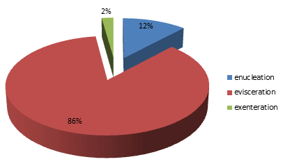
Over the 10 year study period, a total of 4562 new patients had surgeries of various types. Of these, a total of 226 eyes from 226 patients representing 5% (p=0.001*, CI=1.04-1.06) had surgical removal of the eye. The types of eye removal are shown (Fig1).
All excisions were monocular and were of eyes which were considered blind with corrected visual acuity of less than 3/60. The right and left eyes were affected in proportions of 108 and 118, respectively (p=0.51, CI=1.41-1.54).

Figure 1: Distribution of types of surgical removal of the eye.
Age and sex distribution
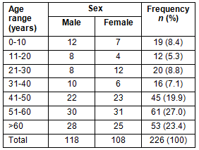
Cohort age and sex distribution is provided (Table 1). Notably, there were more males (n=118, 52.2%) than females (n=108, 47.8%) with a ratio of 1.1:1 (p= 0.50, CI=1.41-1.54). The mean age of the cohort was 47.6±20.2 years (range 2-82). The peak age group was 51-60 years (27%). Children aged under 10 years constituted 8.4%, among whom 4.9% were under the age of 5 years. However, elderly patients (>60 years) constituted 23.4%.
Table 1: Patients' age and sex distribution

Indications for surgical removal of the eyes
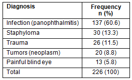
The indications for eye removal are shown (Table 2). Panophthalmitis characterized by gross infection of the uvea and sclera was the commonest cause of eye removal (n=137, 60.6%), followed by staphyloma (13.3%), trauma (11.5%) and then neoplasm (8.7%). Of the 137 cases of panophthalmitis, 3 (2.2%) were post-surgical.
Traumatic cases were characterized by extensive injuries to the globe that were potentially dangerous to be preserved due to sympathetic ophthalmia and cosmetic outlook. Included here were cases of globe rupture, laceration and perforation. Causes of ocular injuries were farm-related (62%), road traffic accidents (14%), assault (12%) and others (12%).
Twenty eyes were removed due to tumors. Of these 14 (70%) of tumors were retinoblastoma, 3 (15%) squamous cell carcinoma of the conjunctiva and 3 (15%) due to non-specific orbital malignancy. All the cases of retinoblastoma were confirmed by histologic evaluation.
Late presentation of patients was common with 69.7% presenting 2 weeks after the onset of symptoms.
Table 2: Indications for eye removal

Discussion
In this study, there were slightly more males, with a male to female ratio of 1.1:1. This is similar to other studies in Turkey, Ethiopia, Southwest Nigeria and South-east Nigeria4,11,13,14. This could be due to males' involvement in high risk activities that predispose them to injuries to ocular structures.
The age group 51-60 years was most frequently affected (27%) and this is higher than in other studies in urban South-east Nigeria11 and Cameroun15. The difference may be due to differences in study area and type of healthcare facility. While the South-east Nigeria study was from an urban tertiary healthcare facility, the Cameroun study was conducted in a pediatric specialty hospital where the most frequently affected age group was 0-10 years. It is common in rural Nigeria to find a preponderance of elderly people because young people are likely to migrate to urban areas in search of corporate employment. In addition, the age group 51-60 years is considered an active age group in rural Nigeria, with many involved in agricultural work and so at risk of farm-related ocular health problems. In this study farm-related ocular injuries were responsible for 62% of all ocular trauma.
Types of surgery
Evisceration was the most common method of eye removal, accounting for 85.8% (n=194) of all destructive eye procedures performed (Fig1). This was followed by enucleation 11.9% (n=27) and exenteration 2.2% (n=5). This is in keeping with a global trend in developing countries where evisceration is more likely to be performed for the more common scenario of ocular trauma and infection16.
Causes of eye removal
Severe eye infection (panophthalmitis): The leading cause of eye removal in this study was panophthalmitis, accounting for 60.6% of cases (Table 2). The dominant causative role of severe eye infection in the indication of eye removal has been reported5,15,17-19; however, the proportion of patients presenting with panophthalmitis was higher in this study. This is a reflection of the poor socioeconomic environment in which these rural communities thrive. Rural dwellers are more likely to have reduced access to modern hospital eye-care services occasioned by unavailability of transportation and poverty. This hospital lacks adequate laboratory services required for effective management of microbial keratitis, which was the dominant primary etiology of panophthalmitis in this study. Overall, there is increased use of traditional eye medication in rural areas, which tends to increase the prevalence of infection-related destructive eye procedures due to its potentially toxic effects and promotion of late presentation. In a similar rural Indian study, Prajna et al reported that 47.7% of the patients with corneal ulcer had applied traditional eye medication prior to presentation at the eye center20. This is also likely in this part of Nigeria.
Staphyloma: Staphyloma was the second highest cause of eye removal (13.3% of all cases). The underlying cause was often not evident, hence inclusion in this group. Other studies5,13,14,21,22 have reported varying significance of staphyloma in eye removal. This finding, however, differed from that in other studies in South-eastern Nigeria by Nwosu7 and Eze et al11, where staphyloma did not result in any eye removal. Both studies were performed at urban tertiary eye centers, while the present study was from a rural primary care center. The higher acceptance of surgical eye removal for cosmetic purposes in the present cohort may be due to differences in the cultural beliefs of those in Ohaozara and its environs. While the predominant Ibos of South-eastern Nigeria believed that removal of the eye would lead to the recurrence of the defect during reincarnation11, the Ohaozara Ibos believe that severe ipsilateral eye problems will involve the contralateral good eye if not removed.
Tumors: Orbitoocular malignancies were the indication for eye removal in 8.9% of cases. Three (15%) were due to squamous cell carcinoma (a marker for HIV infection). There did not appear to be a trend in the occurrence of this tumor over the study period, with one case seen each in 2004, 2007 and 2011. Consistent with the finding in other series4,5,9,11,15, in the present study histologically confirmed retinoblastoma was the commonest tumor type in the pediatric age group, accounting for 100% of all the tumors in those 0-15 years. For all these patients enucleation was the primary form of treatment. This is due to late presentation and the unavailability of alternative modern treatment modalities such as radiotherapy, cytotoxic drugs and laser treatment, which may be necessary for the few patients who presented early. In addition, the effect of poverty tends to negatively affect rural patients' willingness to be referred to modern urban facilities. In a study by Adeoye and Onakpoya, 6 out of the 92 (6.5%) patients studied were unfortunately blind in the second eye4 and this was largely preventable. The high person-blind years and psychosocial implications associated with pediatric sight loss cannot be overstated5. The high incidence among such a vulnerable group is a reflection of the lack of comprehensive capacity to manage such cases in developing countries.
Painful blind eye: In this study, 5.8% of cases of eye removal were due to painful blind eye. Most cases were due to absolute glaucoma. This is less than that in Eze's cohort (13.5%)11 but higher than Olurin's (0.84%), Dawodu and Faal (5.56%) and Vemuganti et al's (3%)6,19,23. Severe ocular pain is an intolerable condition resulting from various ocular diseases and can be treated with topical steroids, cycloplegics, ocular hypotensives, bandage contact lens and retrobulbar alcohol injection24,25. However, if pain is unresolved with treatment, then the eye is surgically removed. Only 5 (38.5%) had received previous treatment with retrobulbar alcohol. The remaining 7 (62.5%) had primary removal of the eye by choice. There was no noticeable change in the incidence of removal of the eye following painful blind eye over the 10 year study period. Significant in this study area is the cultural belief that one sick eye will damage the other good eye. This explains the significant acceptance rate of eye removal following painful blind eye.
Limitations
This study was retrospective and hospital-based and therefore has limited generalizability.
The commonest reason for removal of the eye in rural eye-care practice South-east Nigeria is severe ocular infection (panophthalmitis). This is followed by degenerative globe disorders (staphyloma), and severe eye trauma. More males than females had their eyes surgically removed. Over half of those who had their eyes removed were aged 51 to over 60 years. The commonest cause of eye removal among the children was retinoblastoma. These causes are largely preventable and avoidable. Provision of a system that enhances access to modern eye care at all levels is important. Education of the rural populace on the hazards of ocular injuries, the need for early presentation and avoidance of traditional medication/self-medication will stem the trend of eye removal in rural Nigeria.
Acknowledgement
The medical director of Presbyterian hospital Ohaozara Dr Egwu Ebeyi is thanked for his support and assistance. The nurse in charge of records Mrs Nnenna Onyeabor is appreciated for assisting in retrieving patients' medical records.
References
1. Migliori ME. Enucleation versus evisceration. Current Opinion in Ophthalmology 2002; 13(5): 218-302.
2. Moshfeghi DM, Moshfeghi AA, Finger PT. Major review: Enucleation. Survey of Ophthalmology 2000; 44(4): 277-301.
3. Laura TP, Thomas NH, Timothy JM. Evisceration in the Modern Age. Middle East African Journal of Ophthalmology 2012; 19: 24-33.
4. Adeoye AO, Onakpoya OH. Indications for eye removal in Ile-Ife, Nigeria. African Journal of Medical Science 2007; 36(4): 371-375.
5. Gyasi ME, Amoaku WM, Adjuik M. Causes and Incidence of Destructive Eye Procedures in North-Eastern Ghana. Ghana Medical Journal 2009; 43(3): 122-126.
6. Olurin O. Causes of enucleation in Nigeria. American Journal of Ophthalmology 1973; 73: 987-991.
7. Nwosu SN. Destructive ophthalmic surgical procedures in Onitsha, Nigeria. Nigerian Postgraduate Medical Journal 2005; 12(1): 53-56.
8. Baiyeroju-Agbeja AM, Ajibode HA. Causes of removal of the eye in Ibadan. Nigerian Journal of Surgery 1996; 3: 33-40.
9. Majekodunmi S. Causes of enucleation of the eye at Lagos University Teaching Hospital: A study of 101 eyes. West African Journal of Medicine 1989; 8: 288-291.
10. Onwasigwe EN. The practice of exenteration in Nigeria. Orient Journal of Medicine 2001; 3(3&4): 46-48.
11. Eze BI, Maduka-Okafor FC, Okoye OI, Okoye O. Surgical indication for Eye Removal in Enugu, South Eastern Nigeria. Nigerian Journal of Ophthalmology 2007; 15(2): 44-48.
12. Rourke J. WHO Recommendations to improve retention of rural and remote health workers- important for all countries. Rural and Remote Health 10(4): 1654. (Online) 2010. Available: www.rrh.org.au (Accessed 20 March 2013).
13. Haile M, Alemayehu W. Causes of removal of the eye in Ethiopia. East African Medical Journal 1995; 72(11): 735-738.
14. Gunalp I, Gunduz K, Ozkan M. Causes of enucleation: a clinicopathological study. European Journal of Ophthalmology 1997; 7(3): 223-228.
15. Andre OE, Viola AD, Godfrey K, Abdouramani O, Assumpta LB, Come EM. Indication for destructive eye surgeries at the Yaoundé Gynaeco-Obstetric and Paediatric Hospital. Clinical Ophthalmology 2011; 5: 561-565.
16. Shapiro A, Monselise MB. Destructive ophthalmic procedures, a comparison between a developed and a developing country. Albrecht Von Graefes. Archive for Clinical and Experimental Ophthalmology 1978; 207(4): 271-273.
17. Pandey PR. A profile of destructive surgery in Nepal Eye Hospital. Kathmandu University Medical Journal 2006; 4(1): 65-69.
18. Dada T, Ray M, Tandon R, Vaipayee RB. A study of the indications and changing trends of evisceration in north India. Clinical and experimental Ophthalmology 2002; 30(2): 120-123.
19. Dawodu AO, Faal HB. Enucleation and evisceration in the Gambia. Nigeria Journal of Ophthalmology 2000; 8(1): 29-33.
20. Prajna NV, Pillai MR, Manimegalai TK, Srinivasan M. Use of Traditional Eye Medicines by corneal ulcer patients presenting to a hospital in south India. Indian Journal of Ophthalmology 1999; 47(1): 15-18.
21. Boguseviciene R. An eleven-year experience of eye enucleation caused by severe ocular injuries. Medicina 2005; 41(5): 375-381.
22. Obuchowska I, Sherkawey N, Elmdhms S, Mariak Z, Stankiewiez A. Clinical indications for enucleation in the materials of department of Ophthalmology, Medical Academy in Bialystok in the years 1982-2002. Klinika Oczna 2005; 107(1-3): 75-79.
23. Vemuganti GK, Jalali S, Honavar SG, Shekar GC. Enucleation in a tertiary eye care center in India: Prevalence, current indications and clinicopathological correlation. Eye(London) 2001; 15(6): 760-765.
24. Shah-Desai SD, Tyers AG, Manners RM. Painful blind eye: efficacy of enucleation and evisceration in resolving ocular pain. British Journal of Ophthalmology 2000; 84: 437-438.
25. Al-Faran MF, Al-Omar OM. Retrobulbar alcohol injection in blind painful eyes. Annals of Ophthalmology 1990; 22: 460-462.


